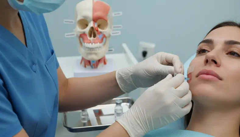
The success of botulinum toxin therapy for temporomandibular disorders depends heavily on proper patient selection. Not every patient presenting with jaw pain is an appropriate candidate for Botox injections, and understanding which patients will benefit most—and which may not respond at all—is critical for achieving consistent clinical outcomes and maintaining patient trust. This article provides a systematic framework for evaluating and selecting TMD patients for neurotoxin therapy.
Diagnostic Criteria for TMD
The Diagnostic Criteria for Temporomandibular Disorders (DC/TMD), published in 2014, provides the current gold standard classification system for TMD diagnosis. Understanding this system is essential for proper patient selection because different TMD subtypes respond differently to neurotoxin therapy.
The DC/TMD classifies TMD into two primary axes. Axis I addresses the physical diagnosis and includes three main categories:
- Myofascial pain (Group I) — Pain originating from the muscles of mastication, including myalgia, myofascial pain with referral, and myofascial pain with spreading. These patients are the best candidates for Botox therapy.
- Disc displacements (Group II) — Including disc displacement with reduction, disc displacement with reduction with intermittent locking, and disc displacement without reduction. These patients may have a secondary muscle component that benefits from Botox but typically require additional interventions.
- Arthralgia/Arthritis/Arthrosis (Group III) — Degenerative joint disease, inflammatory arthritis, and joint pain. These conditions are primarily arthrogenic and are less responsive to neurotoxin therapy targeting the muscles.
Axis II addresses psychological and psychosocial factors, including pain-related disability, depression, anxiety, and jaw functional limitations. Patients with significant Axis II components may require a multidisciplinary approach that includes behavioral health support alongside any neurotoxin therapy.
Imaging Requirements
While imaging is not always required before initiating Botox therapy for myofascial TMD, it serves important roles in ruling out pathology, confirming the diagnosis, and establishing a baseline for treatment monitoring. The imaging modalities most relevant to TMD evaluation include:
Panoramic Radiography
A panoramic radiograph (orthopantomogram) is the minimum imaging standard for initial TMD evaluation. It provides an overview of the condylar morphology, detects gross osseous abnormalities (such as condylar flattening, erosion, osteophyte formation, or fracture), and helps rule out odontogenic sources of pain that may mimic TMD. While panoramic imaging has limited sensitivity for early degenerative changes, it is widely available, cost-effective, and provides sufficient information for most initial assessments.
Cone Beam CT (CBCT)
CBCT provides three-dimensional evaluation of the osseous components of the TMJ and is indicated when panoramic radiography suggests abnormalities or when the clinical presentation warrants detailed condylar assessment. CBCT excels at detecting cortical erosion, subcortical cysts, condylar hyperplasia, and the extent of degenerative joint disease. It is particularly valuable for patients with limited opening, joint crepitus, or progressive changes in occlusion that suggest active articular pathology.
MRI
Magnetic resonance imaging is the gold standard for evaluating soft tissue TMJ structures, including the articular disc, retrodiscal tissue, joint effusion, and surrounding musculature. MRI is indicated when disc displacement is suspected (clicking, locking, sudden changes in occlusion) or when the clinical picture suggests inflammatory or neoplastic pathology. While MRI is not routinely required for patients with straightforward myofascial TMD, it provides critical diagnostic information in complex or atypical cases.
Contraindications
Several absolute and relative contraindications must be evaluated before proceeding with TMJ Botox therapy:
Absolute Contraindications
- Known hypersensitivity to botulinum toxin or any formulation component (human albumin, sodium chloride)
- Infection at the injection site — Active skin infection or abscess overlying the masseter or temporalis
- Neuromuscular disorders — Myasthenia gravis, Lambert-Eaton syndrome, or amyotrophic lateral sclerosis, as botulinum toxin may exacerbate muscle weakness
- Pregnancy and breastfeeding — Botulinum toxin is classified as Category C; insufficient data to establish safety in pregnant or nursing patients
Relative Contraindications
- Aminoglycoside antibiotics — May potentiate the neuromuscular blocking effects of botulinum toxin
- Anticoagulant therapy — Increased risk of bruising at injection sites; consider timing relative to medication schedule
- Unrealistic patient expectations — Patients seeking complete and permanent pain elimination may be disappointed with the temporary and partial relief that neurotoxin therapy provides
- Severe masseter atrophy — Patients who have already undergone extensive muscle reduction may not benefit from further weakening of the muscles of mastication
Identifying Ideal Candidates
The ideal candidate for TMJ Botox therapy presents with a clear myofascial pain component as the primary or dominant feature of their TMD. The following clinical profile characterizes the patients most likely to achieve significant benefit:
Primary myofascial pain: The patient's chief complaint is muscle pain in the masseters, temporalis, or both, rather than joint clicking, locking, or intra-articular pain. Palpation of the muscles reproduces the patient's familiar pain pattern, and the pain is bilateral or unilateral with referral patterns consistent with myofascial trigger points.
Documented bruxism: Evidence of nocturnal or diurnal clenching and grinding, including tooth wear facets, fractured restorations, scalloped tongue margins, buccal ridging (linea alba), and masseter hypertrophy visible on facial examination. Patients who are aware of their clenching habit and can describe jaw fatigue upon waking are particularly good candidates.
Incomplete response to conservative therapy: The patient has attempted and failed or achieved only partial relief from first-line treatments including occlusal splint therapy, physical therapy, NSAIDs, muscle relaxants, and behavioral modification (stress management, habit reversal). Botox is most appropriately positioned as a second-line or adjunctive therapy rather than a first-line treatment.
Clinical Assessment Tools
A standardized clinical assessment protocol ensures consistent patient evaluation and provides objective baseline measurements for tracking treatment outcomes. The following tools and measurements should be part of every TMD evaluation:
- Maximum interincisal opening (MIO): Measured in millimeters using a ruler or Therabite scale. Normal MIO is 40-55mm; values below 35mm suggest restricted opening that may be muscular or articular in origin
- Pain pressure threshold (PPT): Measured using an algometer applied to standardized palpation points on the masseter and temporalis muscles. PPT provides an objective, reproducible measure of muscle tenderness
- Visual analog scale (VAS) or numeric rating scale (NRS): Patient self-reported pain intensity on a 0-10 scale, recorded for current pain, average pain over the past week, and worst pain over the past week
- Jaw Functional Limitation Scale (JFLS): A validated 20-item questionnaire that assesses functional limitations in mastication, jaw mobility, and verbal and emotional expression
- Muscle palpation examination: Systematic bilateral palpation of the masseter (superficial and deep heads), temporalis (anterior, middle, and posterior fibers), medial pterygoid, and lateral pterygoid, recording tenderness levels at each site
Documentation Requirements
Thorough documentation is essential for both clinical care and insurance reimbursement. Every TMJ Botox treatment record should include the following elements:
- Comprehensive history of the TMD complaint, including onset, duration, aggravating and relieving factors, and previous treatments attempted
- Results of the clinical examination, including MIO measurements, muscle palpation findings, joint auscultation, and range-of-motion assessment
- Imaging results and interpretation
- DC/TMD diagnosis with specific subtype classification
- Documentation of failed conservative treatments and rationale for neurotoxin therapy
- Informed consent documenting the risks, benefits, alternatives, and expected outcomes
- Treatment record including the specific botulinum toxin product, lot number, total units administered, injection sites, and any immediate adverse events
- Follow-up plan and outcome measurements at subsequent visits
Our TMJ Injection Therapy Course provides detailed instruction in patient assessment, documentation protocols, and treatment planning for neurotoxin therapy in TMD patients. For practitioners seeking advanced training in complex TMD cases, our Advanced TMJ Treatment Training covers multimodal treatment strategies and challenging clinical scenarios.