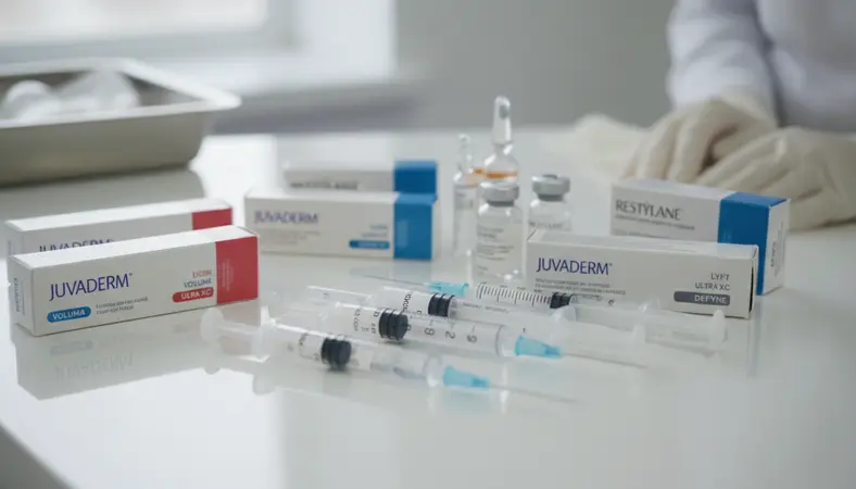
Dermal filler injections are among the most commonly performed aesthetic procedures worldwide, with more than 4.5 million treatments administered annually in the United States alone. When performed by trained practitioners using proper technique, dermal fillers have an excellent safety profile and produce predictable, satisfying results. However, complications can and do occur, and every injector must be prepared to recognize, prevent, and manage them effectively.
This guide provides a comprehensive overview of dermal filler complications, from common and expected side effects to rare but serious adverse events. Understanding this spectrum is not meant to discourage practitioners from offering filler treatments. Rather, it is meant to ensure that every practitioner approaches filler injection with the knowledge and preparation that patient safety demands.
Less than 1%
Incidence rate of serious complications from FDA-approved dermal fillers when performed by properly trained practitioners with thorough anatomical knowledge.
Source: Aesthetic Surgery Journal, 2013
Vascular Occlusion: The Most Serious Complication
Vascular occlusion occurs when filler material is inadvertently injected into or compresses a blood vessel, blocking blood flow to downstream tissue. This is the most feared complication in filler injection because it can lead to tissue necrosis (skin death) if not recognized and treated promptly. In the most severe cases, vascular occlusion involving the ophthalmic artery system can cause partial or complete vision loss, a devastating and potentially irreversible outcome.
The warning signs of vascular occlusion typically appear during or immediately after injection. These include sudden, severe pain disproportionate to the injection itself, blanching (whitening) of the skin in a vascular distribution pattern, dusky or mottled discoloration of the skin, and in cases involving the retinal vasculature, visual changes such as blurred vision, visual field loss, or sudden blindness in one eye. Any of these signs should trigger immediate emergency intervention.
The areas of highest vascular risk include the glabella (between the eyebrows), the nasolabial folds, the nasal dorsum, and the perioral region. These areas contain critical arterial anastomoses that connect to the ophthalmic arterial system. The supratrochlear artery, dorsal nasal artery, angular artery, and superior labial artery are the most commonly involved vessels in occlusion events.
Emergency management of suspected vascular occlusion with hyaluronic acid fillers involves immediate injection of hyaluronidase, the enzyme that dissolves hyaluronic acid. High-dose hyaluronidase (150 to 300 units or more) should be injected directly into and around the affected area to dissolve the obstructing filler material. Additional measures include warm compresses to promote vasodilation, topical nitroglycerin paste to dilate blood vessels, low-dose aspirin to prevent secondary thrombosis, and urgent referral to an ophthalmologist if any visual symptoms are present. Every injection room should have a vascular occlusion emergency kit readily accessible.
Infection
Infection following dermal filler injection is uncommon when proper aseptic technique is maintained, but it can occur and ranges in severity from localized cellulitis to abscess formation to, in rare cases, biofilm infection. Understanding the different presentations and appropriate treatments is essential for every injector.
Acute bacterial infections typically present within days of injection with classic signs of inflammation: increasing redness, warmth, swelling, pain, and sometimes fever. The most common causative organisms are Staphylococcus aureus and Streptococcus species, which can be introduced through the skin puncture during injection or from the patient's own skin flora. Treatment involves broad-spectrum oral antibiotics, with culture and sensitivity testing guiding targeted antibiotic selection if the infection does not respond to empiric therapy.
Biofilm infections present differently and are more challenging to manage. Biofilms are organized communities of bacteria that adhere to the filler material and produce a protective matrix that shields them from antibiotics and the immune system. Biofilm infections often present weeks to months after injection with low-grade swelling, firmness, and tenderness that does not fully resolve with standard antibiotic courses. Management of suspected biofilm requires prolonged antibiotic therapy (often 4 to 6 weeks), hyaluronidase injection to dissolve the filler scaffold supporting the biofilm, and sometimes incision and drainage or filler removal.
Prevention is the best strategy. Maintain strict aseptic technique: clean the injection site with chlorhexidine or alcohol, allow it to dry completely, use sterile product and needles, and avoid injecting through active acne lesions, cold sores, or areas of skin infection. Educate patients about post-treatment infection signs and instruct them to contact the office immediately if symptoms develop.
Granulomas and Foreign Body Reactions
Granulomas are organized inflammatory nodules that form as the body's immune system mounts a response against the injected filler material, which it perceives as a foreign body. While any filler material can theoretically provoke a granulomatous reaction, the risk is significantly higher with permanent fillers like polymethylmethacrylate (PMMA) and silicone compared to temporary hyaluronic acid fillers.
Granulomas typically present months to years after injection as firm, sometimes tender nodules in or near the treatment area. They may be skin-colored or slightly erythematous and can be solitary or multiple. Histologically, they consist of aggregations of macrophages, giant cells, and lymphocytes surrounding the filler material, representing a chronic inflammatory response that the body cannot resolve on its own.
Treatment options for filler granulomas include intralesional corticosteroid injection (triamcinolone acetonide), which suppresses the inflammatory response and can reduce nodule size significantly. For hyaluronic acid fillers, hyaluronidase injection can dissolve the underlying filler and resolve the granuloma. 5-fluorouracil (5-FU) injection has also shown effectiveness for treatment-resistant granulomas. Surgical excision is reserved for cases that do not respond to conservative measures.
Filler Migration
Filler migration refers to the movement of injected filler material away from the intended treatment site. This can result in asymmetry, unnatural contours, or visible lumps in areas adjacent to the injection site. Migration is more common with certain injection techniques, product types, and treatment areas than others.
Factors that increase migration risk include injecting excessive volumes in a single session, using products with lower viscosity or cohesivity than appropriate for the treatment area, injecting into highly mobile areas like the lips without adequate technique to contain the product, and patient-related factors such as massage or manipulation of the treated area too soon after injection.
Lip filler migration, sometimes called "filler shelf" or "filler mustache," has become one of the most discussed complications in aesthetic practice. It occurs when filler injected into the lip body migrates superiorly above the vermilion border, creating a visible ridge or shelf-like appearance. Prevention involves careful injection technique that places product within the appropriate tissue plane, avoiding overfilling, and using products with appropriate rheological properties for lip augmentation.
When migration occurs with hyaluronic acid fillers, targeted hyaluronidase injection can dissolve the misplaced product and restore normal contours. For non-hyaluronic acid fillers, management options are more limited and may require surgical excision in severe cases.
Asymmetry and Aesthetic Complications
Asymmetry is one of the most common patient complaints following filler treatment, though it is important to distinguish between true treatment-induced asymmetry and pre-existing facial asymmetry that becomes more noticeable to the patient after treatment. Pre-treatment photography from multiple angles and thorough consultation about baseline asymmetry can help manage expectations and prevent dissatisfaction.
Treatment-induced asymmetry results from unequal product placement between the two sides of the face. Contributing factors include inconsistent injection depth, uneven product volumes, different tissue responses to the same product volume, and patient factors such as differing muscle activity on each side. Experienced injectors mitigate asymmetry by stepping back frequently during treatment to assess symmetry, comparing both sides from the patient's perspective, and maintaining precise records of product placement and volume.
Other aesthetic complications include overcorrection (too much filler), undercorrection (insufficient filler), the Tyndall effect (a bluish discoloration caused by superficially placed hyaluronic acid filler), and visible or palpable nodules from uneven product distribution. Most of these can be prevented through meticulous technique, appropriate product selection, and conservative dosing with planned touch-up sessions rather than attempting full correction in a single visit.
Prevention Strategies
The most effective approach to filler complications is prevention. While no injection technique can eliminate all risk, adherence to evidence-based prevention strategies dramatically reduces the likelihood and severity of adverse events.
Anatomical knowledge is the primary defense against vascular complications. Understanding the course and depth of facial arteries, identifying high-risk zones, and recognizing anatomical variations between patients are fundamental competencies that can only be developed through comprehensive anatomy training. Never inject in an area where you are uncertain about the vascular anatomy.
Technique modifications that reduce vascular risk include using blunt-tip cannulas instead of sharp needles in high-risk areas, aspirating before injection (though aspiration has limitations and is not foolproof), injecting slowly with small boluses rather than large-volume injections, and maintaining low injection pressure. The "move and inject" technique, where the needle or cannula is repositioned before each small bolus, distributes product more evenly and reduces the risk of large-volume intravascular injection.
Product selection matters. Use the lowest volume of the most appropriate product for each treatment area. Higher-viscosity products with greater cohesivity tend to stay in place better and migrate less than lower-viscosity alternatives. Reversible products, particularly hyaluronic acid fillers, offer a critical safety advantage: if a complication occurs, the product can be dissolved with hyaluronidase.
Emergency Management Protocols
Every practice that performs filler injections must have written emergency protocols and readily accessible emergency supplies. The time between complication recognition and treatment initiation is the most critical factor in outcome, particularly for vascular events. You cannot afford to waste time searching for supplies or looking up protocols when a patient is showing signs of vascular compromise.
Your emergency kit should include hyaluronidase (at least 1,500 units readily available), 2% nitroglycerin paste, warm compresses, aspirin 325mg, and contact information for the nearest ophthalmologist and emergency department. Every staff member who participates in injectable treatments should know where the emergency kit is located and be familiar with the emergency protocol.
Complication management training should be a mandatory component of every injector's education, not an afterthought. The ability to recognize and respond to complications quickly and correctly is what separates a dangerous situation from a manageable one. Our advanced training courses include dedicated complication recognition and management modules with hands-on simulation scenarios that prepare you for real-world emergencies.
Understanding filler complications is not about fostering fear. It is about building the competence and preparedness that allow you to inject with confidence, knowing that you can handle whatever arises. The safest injector is not the one who has never seen a complication but the one who recognizes complications instantly and responds decisively.
Frequently Asked Questions
How common are serious dermal filler complications?
Serious complications such as vascular occlusion are rare, estimated at approximately 1 in 10,000 to 1 in 100,000 injections depending on the treatment area and technique used. Minor complications like bruising, swelling, and temporary asymmetry are much more common, occurring in a significant percentage of treatments. Proper training and technique dramatically reduce the risk of serious adverse events.
Can dermal filler complications be reversed?
Hyaluronic acid fillers, which represent the majority of fillers used today, can be dissolved with the enzyme hyaluronidase. This makes most HA filler complications at least partially reversible. Non-HA fillers like calcium hydroxylapatite and PMMA cannot be enzymatically dissolved, making complication management more challenging for these products.
What should I do if I suspect a vascular occlusion?
Stop injecting immediately. If HA filler was used, inject hyaluronidase directly into and around the affected area at high doses. Apply warm compresses and topical nitroglycerin paste. Administer aspirin. If any visual symptoms are present, arrange immediate ophthalmology evaluation. Document everything and contact your medical director or supervising physician.
How can I minimize the risk of filler complications?
Invest in comprehensive anatomy training, use proper injection techniques including aspiration and slow injection, choose appropriate products for each treatment area, use cannulas in high-risk zones, inject conservatively, maintain aseptic technique, and always have an emergency protocol and supplies readily available. Ongoing education and staying current with best practices are essential.
Should every injector keep hyaluronidase on hand?
Absolutely. Any practitioner who injects hyaluronic acid fillers must have hyaluronidase immediately available in the treatment room. In a vascular emergency, every minute counts. Having to order hyaluronidase or retrieve it from another location could mean the difference between a resolved event and permanent tissue damage or vision loss.