Temporomandibular joint disorder (TMD) affects an estimated 10-15% of the adult population, causing chronic jaw pain, headaches, limited mandibular range of motion, and significant quality-of-life impairment. While traditional treatments such as occlusal splints, physical therapy, and anti-inflammatory medications remain first-line approaches, botulinum toxin (Botox) injections have emerged as a powerful adjunctive and sometimes primary therapy for patients with myofascial-predominant TMD. Understanding the mechanism, dosing protocols, and clinical applications of neurotoxin therapy for TMJ is essential for practitioners offering comprehensive facial pain management.
Mechanism of Action
Botulinum toxin type A works by blocking the release of acetylcholine at the neuromuscular junction, producing a temporary and dose-dependent reduction in muscle contractile force. When injected into the muscles of mastication—primarily the masseter and temporalis—Botox reduces the intensity of involuntary clenching and grinding (bruxism) that drives much of the pain and dysfunction associated with myofascial TMD.
The therapeutic effects of Botox in TMD extend beyond simple muscle relaxation. Research has demonstrated that botulinum toxin also has direct analgesic properties, inhibiting the release of pain-signaling neuropeptides including substance P, calcitonin gene-related peptide (CGRP), and glutamate from peripheral nerve terminals. This dual mechanism—reducing both muscle hyperactivity and nociceptive signaling—explains why many TMD patients experience pain relief that exceeds what would be expected from muscle relaxation alone.
Additionally, by reducing the force generated by the muscles of mastication, Botox decreases the mechanical load on the temporomandibular joint itself, allowing inflamed joint structures to heal and potentially slowing the progression of degenerative joint changes in patients with combined myofascial and arthrogenic TMD.
Dosing Protocols for Masseter and Temporalis
Masseter Muscle
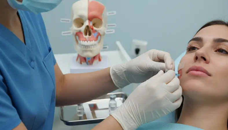
The masseter is the primary target muscle for TMJ Botox therapy. A typical starting dose ranges from 25 to 50 units of onabotulinumtoxinA (Botox) per side, divided among three to four injection points distributed across the body of the muscle. The injection sites should be placed within the inferior two-thirds of the masseter to avoid affecting the parotid gland and zygomatic branch of the facial nerve, which lie superficial to the superior portion of the muscle.
To locate the masseter, ask the patient to clench their teeth firmly and palpate the muscle belly. Mark three to four injection points approximately 1 cm apart, arranged in a triangular or grid pattern within the thickest portion of the muscle. Use a 30-gauge or 32-gauge needle, inserting to a depth of approximately 5-8mm to reach the muscle belly. Each injection point typically receives 5-10 units.
Temporalis Muscle
The temporalis muscle is the second most commonly treated muscle in TMJ Botox therapy. A typical dose ranges from 15 to 30 units per side, divided among three to five injection points along the anterior and middle portions of the muscle. The injection sites should be placed at least 1 cm above the superior temporal line to avoid the temporal branch of the facial nerve.
Palpate the temporalis by asking the patient to clench and release repeatedly while you feel the muscle contract in the temporal fossa. Inject at a shallow depth (3-5mm) to reach the muscle, as the temporalis is relatively thin compared to the masseter. Each injection point typically receives 5 units.
Total Dose and Treatment Schedule
The total dose per session typically ranges from 80 to 160 units when treating both masseters and both temporalis muscles bilaterally. Conservative practitioners may begin with lower doses (80-100 units total) for the initial treatment and increase at subsequent sessions based on clinical response. Treatment intervals are generally every 12-16 weeks, as the effects of botulinum toxin typically last three to four months. Some patients develop longer-lasting muscle atrophy over time, allowing for extended intervals between treatments.
Patient Selection
Not all TMD patients are appropriate candidates for Botox therapy. The best responders are patients with myofascial-predominant TMD characterized by masseter and temporalis hyperactivity, nocturnal or diurnal bruxism, muscle tenderness on palpation, and headaches originating from the temporal region. Patients with purely arthrogenic TMD (disc displacement, osteoarthritis, or joint hypermobility without significant muscle involvement) are less likely to benefit from neurotoxin therapy alone.
Ideal candidates typically present with one or more of the following clinical features:
- Bilateral masseter and/or temporalis pain on palpation
- Visible masseter hypertrophy (squaring of the jaw angle)
- Evidence of bruxism (tooth wear facets, scalloped tongue borders, buccal ridge line)
- Tension-type headaches in the temporal region
- Failure to achieve adequate relief with conservative therapies (splints, physical therapy, NSAIDs)
- Limited mouth opening due to muscle guarding (as opposed to mechanical disc obstruction)
Treatment Timeline and Expectations
Setting appropriate patient expectations is crucial for treatment satisfaction. Botox does not produce immediate pain relief. The onset of neuromuscular blockade typically begins three to five days after injection, with peak effect reached at two to four weeks. Many patients notice a reduction in clenching intensity within the first week, followed by progressive improvement in pain, headaches, and jaw function over the subsequent two to three weeks.
It is important to counsel patients that the first treatment session may not produce the full therapeutic effect. Many practitioners observe that the second and third treatment sessions produce superior and more sustained results, likely due to cumulative muscle atrophy and central nervous system adaptation. A minimum commitment of two to three treatment cycles (6-9 months) is recommended before determining whether a patient is a long-term responder to Botox therapy for TMD.
Combination with Splint Therapy
Botox and occlusal splint therapy are complementary rather than competing treatments. Many TMD specialists employ a combined approach, using Botox to reduce muscle hyperactivity and pain while simultaneously using a stabilization splint to optimize occlusal relationships and protect the dentition from further wear. The synergistic effect of this combination often produces results superior to either treatment alone.
When combining therapies, Botox is typically administered first to reduce muscle tension and pain, making it easier for the patient to tolerate splint fabrication and adjustment. The splint is then delivered one to two weeks after the Botox injection, once the neurotoxin has reached its initial therapeutic effect. This sequencing allows the splint to be adjusted to the patient's reduced muscle force profile, improving both comfort and efficacy.
To develop proficiency in neurotoxin therapy for TMJ disorders, our TMJ Injection Therapy Course and Advanced TMJ Treatment Training provide comprehensive hands-on instruction in injection technique, dosing protocols, patient assessment, and multimodal treatment planning. For practitioners new to neurotoxin injection, our Botox Certification Course offers foundational training in botulinum toxin pharmacology and injection fundamentals.